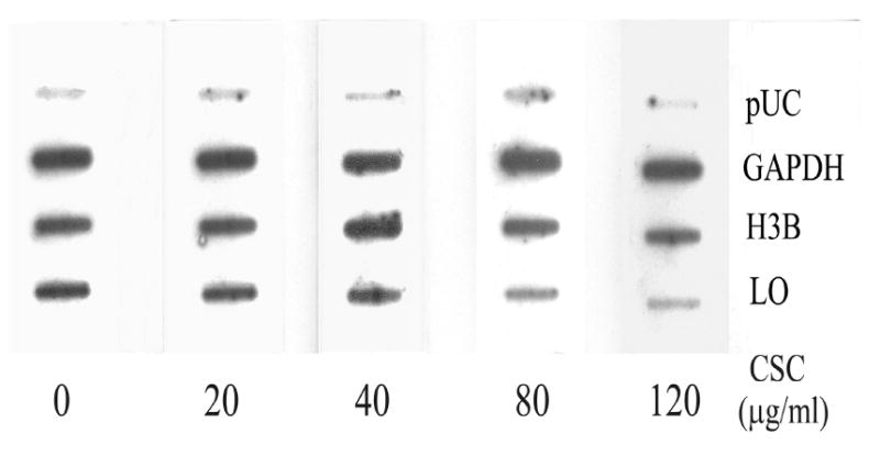
Figure 2. Nuclear run-on assay for relative transcription rates of LO in control and CSC-treated cells.

Nuclei were freshly isolated from control and CSC-treated cells under the same conditions as described in Figure 1. Nascent transcripts were labeled with 32P-UTP and hybridized to a previously prepared filter containing cDNAs for LO, histone 3B (H3B) and glyceraldehydes-3-phosphate dehydrogenase (GAPDH) and pUC plasmids without insert (pUC).
