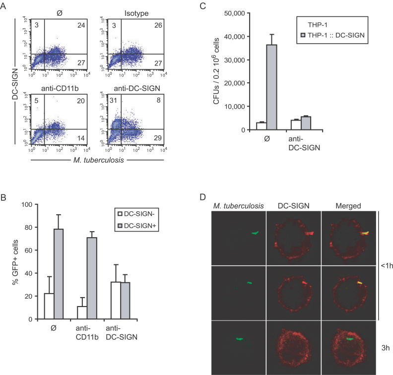
Figure 4. DC-SIGN Mediates M. tuberculosis Binding to Alveolar Mφs from Patients with TB.
(A) Alveolar Mφs from a patient with TB were infected with GFP-expressing M. tuberculosis, in the absence (ø; upper left panel) or the presence of control isotype (upper right panel), anti-CD11b (lower left panel), or -DC-SIGN (lower right panel) blocking antibodies. In the upper panels, cells were then stained with fluorescent PE-conjugated anti-DC-SIGN and APC-conjugated anti-CD11b antibodies. In lower panels, fluorescent antibodies were added together with blocking antibodies (same clones).
(B) Proportion of GFP+ cells in DC-SIGN− (open bars) and DC-SIGN+ (grey bars) alveolar Mφs as calculated from (A) using BALs from two patients with TB. THP1 Mφs expressing or not expressing DC-SIGN (THP1::DC-SIGN) were used in a binding experiment with M. tuberculosis H37Rv, in the presence or absence of anti-DC-SIGN antibodies.
(D) Confocal microscopy examination of adherent DC-SIGN+ cells infected with GFP-expressing M. tuberculosis for various times.

