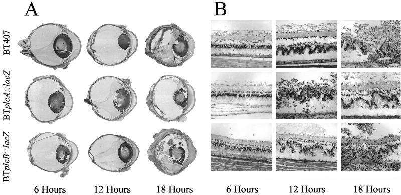
FIG. 7.
Histologic analysis of experimental B. thuringiensis endophthalmitis. Representative whole-organ (A) and retinal (B) histology of eyes intravitreally injected with strains BT407, BTplcA::lacZ, and BTplcB::lacZ at 6, 12, and 18 h postinfection are shown. All histologic sections were stained with hematoxylin and eosin. In whole-organ sections, severe inflammation and retinal detachment were observed by 18 h. Photoreceptor folding and detachment were observed in retinal sections by 12 h. By 18 h, retinal layers were virtually indistinguishable. Gross pathologic changes observed in all infection groups were similar at all time points. Magnifications, ×9 (A) and ×180 (B). (Top row of panel A is reprinted from reference 11a with permission of the publisher.)

