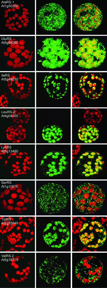
Fig. 1.
Mitochondrial and/or chloroplast import of targeting sequence GFP or RFP fusions in tobacco protoplasts. The images are false-color maximum projections from a confocal microscope. The red channel (Left) shows chlorophyll autofluorescence, the green channel (Middle) shows GFP or RFP fluorescence (RFP for GluRS and ThrRS, GFP for all others); in Right, the two channels are superimposed. Several of the images include untransformed protoplasts in the field of view to demonstrate the lack of background fluorescence under the conditions used. The scale of the images and cell size varies slightly, but the chloroplasts are a uniform 5 μm in these protoplasts.

