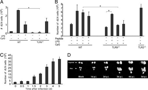
Fig. 1.
TLR agonists alone are sufficient to induce LN hypertrophy. (A) The number of cells in popliteal LN was measured in WT or TLR9-/- mice (n = 4 per condition) 4 days after footpad injection of TLR ligands in saline. (B) The number of cells in popliteal LN was measured in WT, TLR9-/-, or TLR2-/- mice (n = 3 per condition) 4 days after footpad injection of TLR ligands with or without OVA323–339 peptide in IFA. (C) The total cell number of dLNs (inguinal and iliac) after ivag HSV-2 (106 PFU) infection was measured at the indicated time points. (D) Photographic depiction of dLNs of HSV-2-infected mice. Similar results were obtained in five separate experiments. *, P < 0.05, significant difference between the indicated groups.

