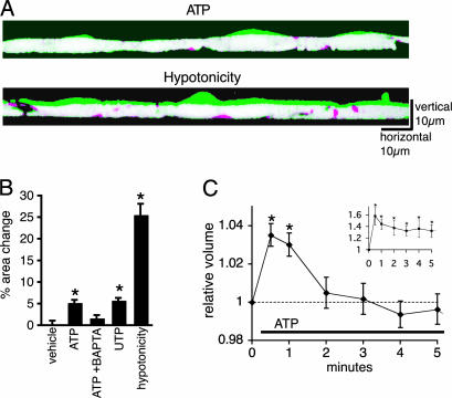
Fig. 1.
Astrocytic Ca2+ increases are associated with a transient increase in cell volume. (A) Exposure to ATP (100 μM) induces swelling of cultured astrocytes. Confocal vertical cross-sectional images of confluent astrocyte cultures with 3-5 cells in the field of view loaded with calcein/AM (5 μM for 30 min) were constructed from repetitive x-z line scans at 488 nm excitation. Two images of cross-sectional area before (red) and 1 min after the exposure to ATP (green) are overlapped to indicate the change in cell volume. Overlapped areas (no change before and after ATP exposure) are displayed as white. Hypotonicity (214 mOsM) also induced cellular swelling. (B) Quantification of relative changes in cross-sectional areas 1 min after addition of vehicle (control, n = 12); ATP (100 μM, n = 23); ATP to cultures preloaded with BAPTA (20 μM for 30 min, n = 11); UTP (100 μM, n = 15) and hypotonicity (214 mOsM, n = 6). *, P < 0.01 compared with control, Tukey-Kramer test. (C) Coulter counter analysis of relative changes in astrocytic cell volume evoked by ATP. ATP exposure of astrocytes in suspension triggered a transient increase in cell volume at 30 and 60 sec. (C Inset) Hypotonicity induced a large and sustained increase in astrocytic cell volume. n = 5; *, P < 0.05 compared with control, t test. mean ± SEM.

