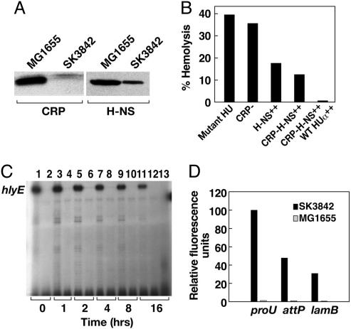
Fig. 6.
Direct influence of HUαE38K,V42L-mediated nucleoid reorganization on specific changes in transcription profile. (A) Western blot analysis of CRP and H-NS concentration in midlog-phase cultures of wild-type and hupA mutant cells. (B) Hemolytic activity of hupA mutant strains with altered levels of transcription regulators for hlyA. From left to right, the strains used were SK3842, SK3842 Δcrp, SK3842 (pHNS42), SK3842 Δcrp (pHNS42), and SK3842 Δcrp (pHNS42 and pHU-GFPuv). Assays were done 16 h after the addition of IPTG. (C) Primer extension analysis of hlyE mRNA expression in SK3842 Δcrp pHNS42 (lanes 1, 3, 5, 7, 9, and 11) and MG1655 Δcrp pHNS42 (lanes 2, 4, 6, 8, 10, and 12) at different time points after induction of H-NS. Lane 13 shows the hlyE mRNA expression in SK3842 Δcrp pHNS42, pHU-GFPuv at 16 h after IPTG addition. (D) GFP fluorescence intensities from PproU–GFP translation fusions at the proU, attP, and lamB chromosomal loci in the wild-type and hupA mutant strains.

