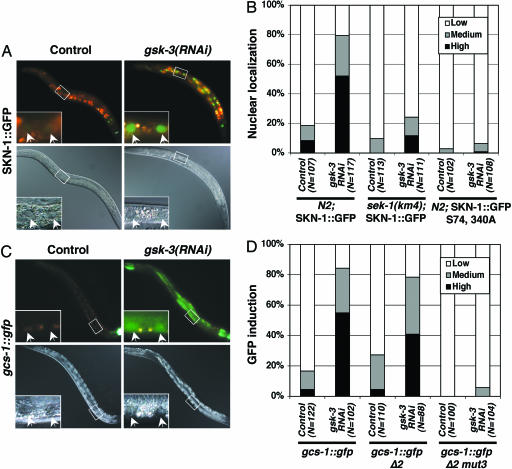
Fig. 1.
gsk-3 inhibits SKN-1 localization and SKN-1 target gene expression. (A and B) SKN-1 is localized to intestinal nuclei in gsk-3(RNAi) animals in the absence of stress. N2 Is007[SKN-1::GFP], sek-1(km4) Is007[SKN-1::GFP], and N2 Ex[SKN-1::GFP S74, 340A] L4 animals were placed onto nematode growth medium plates containing E. coli HT115 that carried either gsk-3 dsRNA or control (L4440) feeding vectors. The animals were then incubated for 24 h at 20°C and allowed to lay eggs. Their surviving progeny were scored as L4 larvae and young adults for accumulation of SKN-1::GFP in intestinal nuclei as described in ref. 11. “High” indicates that a strong SKN-1::GFP signal was present in all intestinal nuclei, as in the gsk-3(RNAi) animal shown. “Medium” refers to animals in which nuclear SKN-1::GFP was present at high levels anteriorly or anteriorly and posteriorly but was barely detectable midway through the intestine. sek-1 encodes a C. elegans p38 MAPK kinase and is required for SKN-1 function in the intestine (see Results and Discussion) (28). (C and D) gcs-1::gfp is expressed constitutively in gsk-3(RNAi) animals. gsk-3 RNAi was performed in gcs-1::gfp, gcs-1::gfp Δ2, and gcs-1::gfp Δ2-mut3 strains as in A and B, then intestinal gcs-1::gfp expression was scored in F1 L4 larvae and young adults (11). “High” indicates that gcs-1::gfp was present at high levels anteriorly and was detectable throughout most of the intestine, as in the gsk-3(RNAi) image shown. “Medium” refers to animals in which gcs-1::gfp was present at high levels anteriorly and possibly posteriorly but was not detected in between. The gcs-1::gfp fluorescence apparent in the control image derives from SKN-1-independent pharyngeal expression (11). In A and C, upper images show fluorescence, lower images show Nomarski imaging, and pairs of intestinal nuclei (arrows) are shown in each inset.

