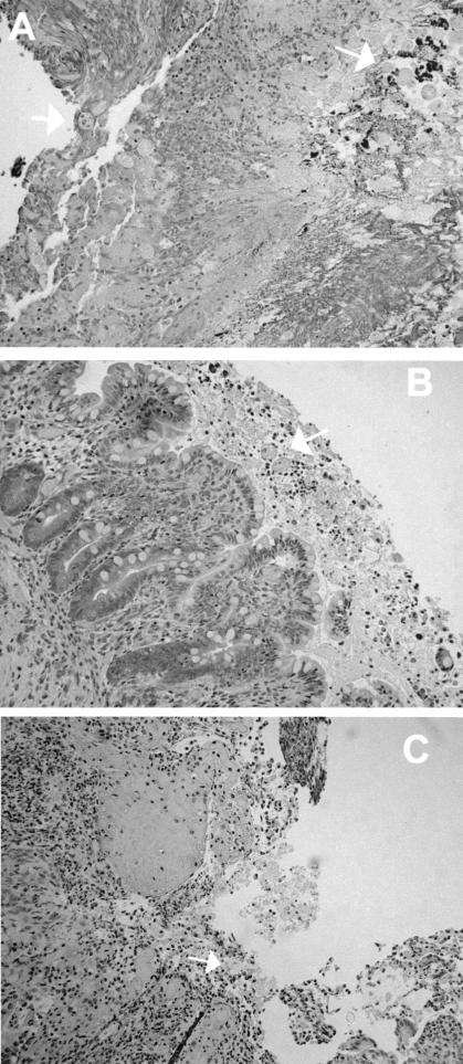
FIG. 2.
Morphologic findings in E. histolytica-infected human colonic xenografts from SCID-HU-INT mice. Photomicrographs of hematoxylin-and-eosin-stained sections of E. histolytica-infected human colonic xenografts from SCID-HU-INT mice treated with 1B10 (A) or EH5 (B and C) are shown. (A) Extensive mucosal destruction and inflammation and multiple E. histolytica trophozoites (white arrows) that have invaded the mucosa and submucosal tissues are visible in a section from a 1B10-treated SCID-HU-INT mouse. (B) In an E. histolytica-infected human colonic xenograft from an EH5-treated SCID-HU-INT mouse, amebic trophozoites are clumped together in the lumen but the mucosa remains intact and no signs of invasion arevisible. (C) In a section from a different EH-5-treated SCID-HU-INT mouse, mucosal destruction and inflammation are present and amebic trophozoites can be seen invading the mucosa and submucosal regions. Magnification, ×176 (all panels).

