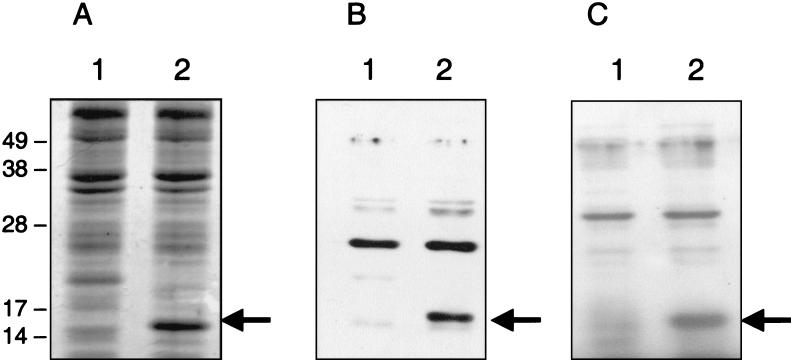
FIG. 8.
SDS-PAGE analysis of crude cell lysates from E. coli strains BL21(pT7-5) (lanes 1) and BL21(pLPFA01) (lanes 2) after overnight incubation with 0.1 M IPTG. The corresponding lysates were separated and either stained with Coomassie brilliant blue (A) or transferred to polyvinylidene difluoride membranes for Western blotting with LpfA peptide antiserum (B) or S. enterica serovar Typhimurium LpfA antiserum (C). The molecular mass markers (in kilodaltons) are indicated on the left, and the 16-kDa EHEC LpfA protein in each gel is identified with an arrow.

