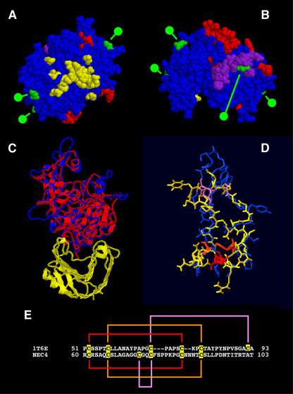
Figure 2.
NEC4 protein models based on the structure of the TAXI-1. A and B, Model based on threading of NEC4 sequence through the coordinates of 1T6E. Residues shown in red represent the loops found in the tobacco NEC4 but not in TAXI-1. Residues shown in green indicate the locations of putative N-glycosylation sites. Shown in yellow are NEC4 residues that are homologous to TAXI-I residues that are within 4 Å of its ligand (an A. niger xylanase) as identified from the model shown in C. Residues shown in purple indicate the location of the pseudoknottin domain. A, Bottom of the NEC4 model looking up from the hemicellulase ligand (assuming that NEC4 binds its XEG ligand with a geometry that is similar to the binding of xylanase by TAXI-I). B, Top of the same NEC4 model. C, Second model based on the replacement of the TAXI-1 molecule of 1T6G with the refined NEC4 model; the ribbon diagram of the A. niger xylanase is in yellow and the ribbon diagram of TAXI-1 is in red. This is overlaid with the NEC4 Cα backbone (in blue). D, Knottin domain of TAXI-1 in blue overlaid with the pseudoknottin domain of NEC4 (in yellow). The three disulfide linkages are color coded (red, orange, and purple). E, Amino acid sequences of the knottin domain of TAXI-1 and the corresponding pseudoknottin domain of NEC4. Cys residues are highlighted and disulfide linkages are color coded to match those shown in D.

