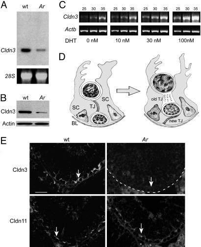
Fig. 1.
Cldn3 expression is regulated by androgens. (A Upper) Northern blot analysis on total RNA from testes of 2-month-old wild-type and Arinvflox(ex1-neo)/Y;Tg (Amh-Cre) mice and probed with a 32P-labeled Cldn3 cDNA fragment. (Lower) 28S rRNA was used as a loading control. (B) Western blot analysis of total testis protein extracts prepared from wild-type and Ar mutant mice probed with anti-Cldn3 antibody (Upper) and an anti-actin antibody as a loading control (Lower). (C) Semiquantitative RT-PCR analysis of Cldn3 expression in immortalized Sertoli-like TM4 cells transiently transfected with a cDNA encoding the androgen receptor. RNA was harvested from cells 48 h after treatment with varying doses of dihydrotestosterone (DHT), reverse transcribed, and amplified by PCR with primers specific for Cldn3 (Upper) or Actb (Lower). Reactions were terminated after 25, 30, and 35 cycles. (D) Schematic drawing of a transverse section through the seminiferous epithelium. The epithelium is segregated into basal and adluminal compartments by the formation of TJs between neighboring Sertoli cells (15). As germ cells move from the basal to the adluminal compartment, new TJs form and old TJs are disassembled. Expression of the androgen receptor in Sertoli cell nuclei is maximal during the stages of new TJ formation (8, 16). SC, Sertoli cell; PL, preleptotene spermatocyte; PS, pachytene spermatocyte; BL, basal lamina. (E) Immunofluorescence detection of Cldn3 and Cldn11 on serial sections from wild-type and Ar mutant testes. Both proteins are localized to TJs in the basal compartment of the seminiferous epithelium, although the staining of Cldn3 (arrow in Upper Left) appears to extend beyond that of the staining of Cldn11 (arrow in Lower Left). Expression of Cldn3 is absent in the region of Sertoli cell TJs in Ar mutants (Upper Right). The basal lamina of the tubule is outlined with a dotted line. Expression of Cldn11 is retained in Ar mutant testis (Lower Right). (Scale bar: 50 μm.)

