Fig. 2.
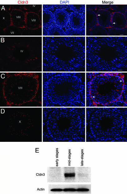
Expression of Cldn3 is restricted to newly formed TJs. (A–D) Immunofluorescence localization of Cldn3 in adult testis. Nuclei were stained with DAPI and the tubules staged based on the particular association of germ cells within a tubule (44). The arrows indicate regions of Cldn3 detection. High magnification images show the absence of Cldn3 in Stages IV and X and its presence in Stage VIII. (Scale bars: A, 50 μm; B, 20 μm for B–D). (E) Western analysis of Cldn3 in extracts prepared from seminiferous tubule fragments identified by transillumination (30). Actin was used as a loading control.

