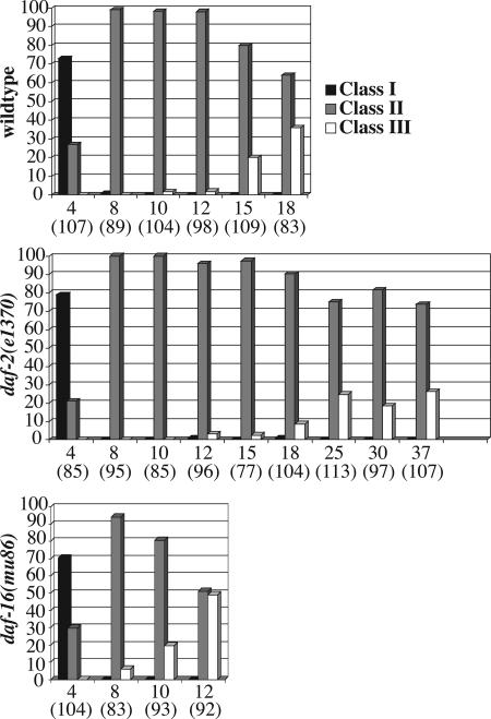
Fig. 2.
Summary of nuclear morphology changes in aging animals of different genetic backgrounds. Nuclear morphology of wild-type, daf-2(e1370), and daf-16(mu86) animals was monitored by using ccIs4810(lmn-1::gfp). Multiple images were taken of the middle part of the worm (excluding the head and the tail) by a confocal or a compound microscope. Nuclei from the collected images were counted and grouped into three different classes (class I–III) based on their morphology and GFP signal distribution (see Results). Neuronal nuclei were excluded from the counting. The nuclei counted are primarily hypodermal. Data were derived from a total of 20–30 animals on each of the days indicated (pooled from two to three experiments). y axis, percent of nuclei in a given category (black column, class I; gray column, class II; white column, class III). x axis, days (total number of nuclei scored).

