Fig. 2.
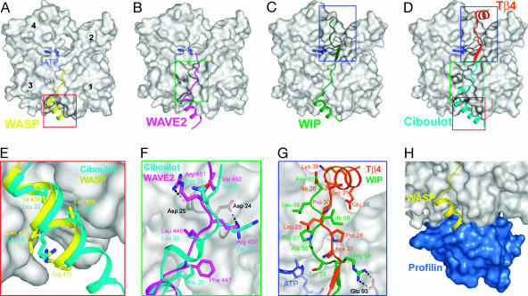
WH2–actin structures. (A–C) Structures of the WH2 domains of WASP, WAVE2, and WIP determined as ternary complexes with actin (gray) and DNase I (see Fig. 5). (D) Superimposition of the structures of ciboulot (9) and Tβ4 (10), which together represent Tβ–actin. (E–G) Close-view comparisons of different parts of the WH2–actin and Tβ–actin structures shown in A–D. (H) Partial overlap between the actin-binding sites of profilin (blue) and WH2.

