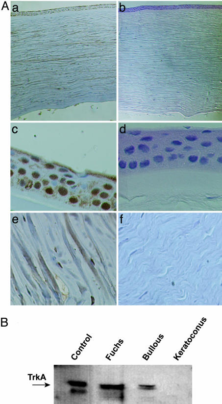
Fig. 3.
Lack of TrkANGFR protein expression in the cornea of keratoconus patients. (A) Representative immunohistochemical staining of TrkANGFR in control (a) and keratoconus (b) corneas. Higher magnification shows the lack of TrkANGFR in corneal cells, including epithelium (compare c with d) and keratocytes (compare e with f). Sections were counterstained with Harri's hematoxylin. (B) Western blot analysis of corneal protein extracts. The immunoreactive levels of TrkANGFR (arrow) are shown for control, Fuchs' dystrophy, bullous, and keratoconus corneas. In the first lane (Control), the lower band is a nonspecific immunoreactive band.

