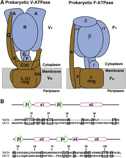
Figure 1.

(A) Schematic models of the prokaryotic V-ATPase from T. thermophilus and the prokaryotic F-ATPase from E. coli. The V1 and F1 domains are colored in blue, and the F0 and V0 domains are in brown. (B) Sequence alignment of subunit F from the prokaryote T. thermophilus and the eukaryotic yeast Saccharomyces cerevisiae, with secondary structure from our T. thermophilus structure. The sequence alignment was performed using program water of EMBOSS (Rice et al, 2000). Identical residues in this alignment are enclosed by thick rectangles. Identity 23.5%, similarity 40.9%, and gaps 18.3% were shown in this alignment. Sequence numbering is for T. thermophilus. THETH, T. thermophilus; SACCE, S. cerevisiae.
