Figure 5.
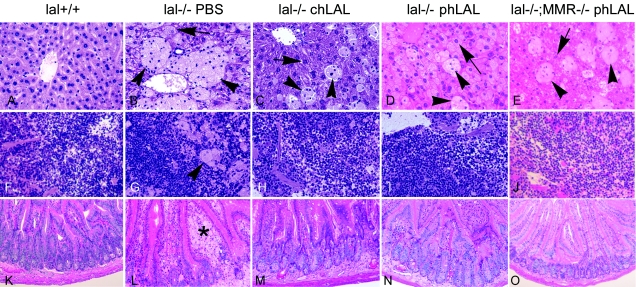
Histological analyses of wild-type, lal−/−;MMR+/+, and lal−/−;MMR−/− mice administered PBS, phLAL, or chLAL. Shown are representative H+E-stained tissue sections from age-matched wild-type (lal+/+) mice (A, F, and K); from lal−/−;MMR+/+ mice injected with PBS (B, G, and L), chLAL (C, H, and M), or phLAL (D, I, and N); and from lal−/−;MMR−/− mice treated with phLAL (E, J, and O). The tissues are liver (A–E), spleen (F–J), and small intestine (K–O). Arrows indicate hepatocytes, arrowheads indicate Kupffer cells, and the asterisk (*) indicates intestinal macrophages. The numbers of remaining Kupffer storage cells in the phLAL- or chLAL-treated mouse livers are similar, whereas the hepatocytes in the chLAL-treated lal−/−;MMR+/+ mice and the phLAL-treated lal−/−;MMR−/− mice are essentially cleared of storage. Hepatocyte storage remained in the phLAL-treated lal−/−;MMR+/+ mice. In spleen and small intestine, the storage macrophages were nearly eliminated in all phLAL-treated mice. Original magnification, ×200.

