
Fig. 1. Normal and cancerous breast morphology.
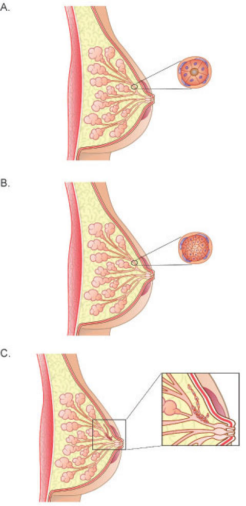
A) Diagram showing the normal organization of ducts and acini in the human breast. The cross-section demonstrates luminal epithelial cells aligned in a polar manner so their apical side faces and surrounds the lumen. The luminal epithelial cells are surrounded by a non-continuous layer of myoepithelial cells. Surrounding these cells is the basement membrane. Fibroblasts align the basement membrane and this entire structure is surrounded by the stroma, which is predominantly, but not exclusively, composed of type I collagen. B) During ductal carcinoma in situ (DCIS), the normal polar organization of the luminal epithelial cells is lost, as these cells de-differentiate and proliferate. The cross-section shows the epithelial cells completely filling the lumen. In less severe cases of DCIS, the luminal epithelial cells do not completely block the lumen. In DCIS, the transformed epithelial cells do not cross the basement membrane, but remain within the duct. C) DCIS sometimes leads to invasive, or infiltrating, carcinoma, in which the epithelial cells migrate and invade through the basement membrane and into the surrounding stroma.