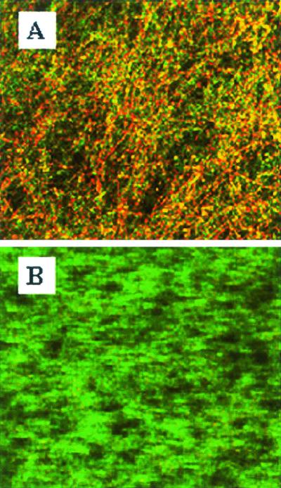
FIG. 3.
CLSM of C. albicans 3153A biofilms on plastic coverslips after incubation for 24 h with medium alone (A) or with 0.5 μg of caspofungin per ml (B). Experiments utilize the FUN 1 stain to directly visualize the effects of caspofungin on preformed biofilms. Note the shift from green to red fluorescence visible in panel A (untreated control biofilms), which reflects processing of the dye by metabolically active cells. In contrast, caspofungin-treated biofilms (B) show diffuse green fluorescence characteristic of dead cells. Images are single xy optical sections taken across the z axis. Magnification, ×200.

