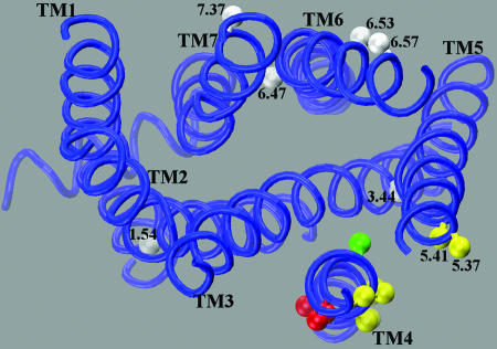
Fig. 4.
Homology model of the D2R based on the structure of bovine rhodopsin (52). The Cβ positions that were crosslinked by CuP are shown in yellow or red depending on their predicted location at the AFM or ECM interfaces (see Fig. 1). Position 4.56, which is predicted to face inward is shown in green. For simplicity, we omit the segment 4.60-4.62, which is the most extracellular part of TM4 and may not be in an α-helical conformation. Endogenous cysteines that did not crosslink, including 1.54, 3.44, and 6.47, are shown in gray, as are the positions of substituted cysteines 6.53, 6.57, and 7.37 in TM6 and TM7 that did not crosslink despite their predicted positions facing outward in the rhodopsin monomer.

