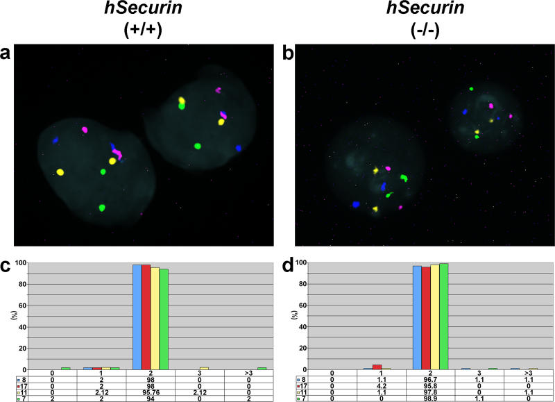
Figure 2. Assessment of Chromosomal Stability in Interphase Nuclei of Parental HCT116 Cells and Chromosomally Stable hSecurin−/− Cells Using Centromere-Specific Probes for Chromosomes 7, 8, 11, and 17.
(A and B) Representative interphase FISH images of parental HCT116 (hSecurin+/+) (A) and hSecurin−/− (B) cell nuclei after hybridization of a four-color probe set consisting of centromere probes for chromosomes 7 (Cy5.5; purple), 8 (FITC; green), 11 (Cy5; blue), and 17 (Cy3; yellow). In each nucleus, two signals are visible for each probe.
(C and D) Graphic summary of chromosome gains and losses in parental HCT116 (C) and hSecurin−/− (D) cells. The percentage of signals per nucleus for chromosomes 7, 8, 11, and 17 was determined from 300 cells of each genotype (100 cells each in three separate experiments).

