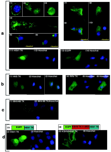
FIG. 2.
EGFP-KSHV TK and related gamma-2 herpesvirus TKs demonstrate a similar pattern of intracellular localization. (a) EGFP-KSHV TK localizes to the cytoplasm: 293 cells (panels 1 to 3), 143B TK- cells (panels 4 to 6), and primary keratinocytes (panels 7 to 10), whereas EGFP-HSV1 TK expressed in 143B TK- cells (panel 11) localizes to the nucleus and EGFP alone (panel 13) localizes diffusely. (b) EGFP-HVS TK (panel 1) and EGFP-RRV TK (panel 3), as well as EGFP-MHV-68 TK (c, panels 1 to 2), all localize to cytoplasmic compartments. (d) EGFP-HSV1 TK (panel 2) localizes to the nucleus; however, the chimera EGFP-KSHV/HSV1 TK (panel 5) localizes to the cytoplasm. Schematic diagrams detailing the structure of EGFP-HSV1 TK and EGFP-KSHV/HSV1 TK are shown in panels 1 and 4, respectively. Cells counterstained with Hoechst for nuclear discrimination are shown in panels 1, 5, 6, 8, 10, 12, and 14 (a), 2 and 4 (b), 2 (c), and 3 and 6 (d) (blue staining).

