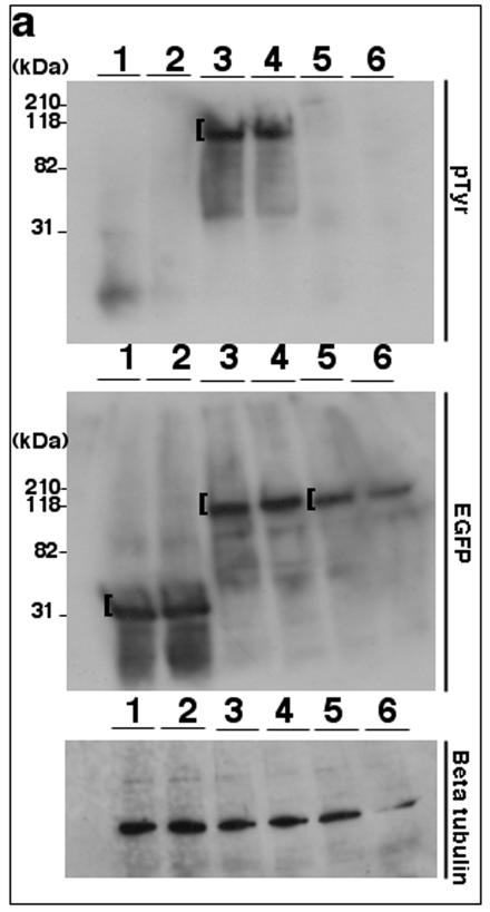
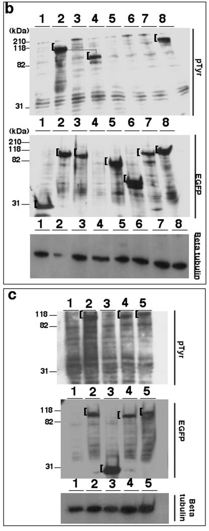
FIG.7.
Tyrosine phosphorylation of TK is conserved among related rhadinoviruses. (a) Lysates from 293T cells (15 μg/lane) expressing EGFP (lanes 1 and 2), EGFP-KSHV TK (lanes 3 and 4), or EGFP-KSHV TKΔATP (lanes 5 and 6) were stained using either an anti-pTyr (top) or an anti-GFP antibody (middle). (b) Equivalent lysates expressing EGFP (lane 1), EGFP-KSHV TK (lane 2), EGFP-KSHV TKΔATP (lane 3), nontagged KSHV TK (lane 4), EGFP-HSV1 TK (lane 5), EGFP-vaccinia virus TK (lane 6), EGFP-EBV TK (lane 7), or EGFP-MHV-68 TK (lane 8) were stained using an anti-pTyr (top) or an anti-GFP monoclonal antibody (middle). (c) Lysates prepared from 293T cells (15 μg) alone (lane 1) or expressing EGFP-KSHV TK (lane 2), EGFP (lane 3), EGFP-HVS TK (lane 4), or EGFP-RRV TK (lane 5) were stained using anti-pTyr (top) or an anti GFP antibody (middle). Protein loading (control) blots were developed using an antibody to β-tubulin and are shown on the bottom of panels a to c. A bracket ([) indicates detected expressed fusion proteins.


