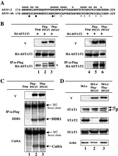
FIG. 1.
hPIV4 V protein complex formation with STAT1, STAT2, DDB1, and Cul4A without STAT degradation. (A) Amino acid sequences of the V-specific regions of hPIV2 and hPIV4A V proteins. The symbols above and below the sequences indicate the positions of conserved cysteine (asterisks), tryptophan (or tyrosine) (squares), and common (circles) residues. (B) Interaction of V proteins with STAT1 or STAT2. BSR T7/5 cells were transfected with either pTM-HA-hSTAT1 (lanes 1 to 3) or pTM-HA-hSTAT2 (lanes 4 to 6) plus pCI carrying the Flag epitope-tagged hPIV2 V gene (lanes 2 and 5), the hPIV4A V gene (lanes 3 and 6), or an empty pCI (lanes 1 and 4). Whole-cell extracts were prepared at 48 h posttransfection, and samples containing equal amounts of total protein were assayed by Western blotting for their levels of HA-hSTAT1 or HA-hSTAT2 and Flag-V proteins using α-HA and α-Flag antibodies, respectively. Other samples were first immunoprecipitated (IP) with Flag affinity gel (Sigma), and the selected material was then assayed by Western blotting for detection of hSTAT proteins. The asterisk on the right indicates the immunoglobulin heavy chain. (C) Interaction of V proteins with DDB1 or Cul4A. HeLa cells were transfected with pCI-Flag-PIV2V (lane 2), pCI-Flag-PIV4V (lane 3), or an empty pCI (lane 1). Whole-cell extracts were prepared at 48 h posttransfection, and samples containing equal amounts of total protein were assayed by Western blotting for detection of Flag-V proteins (α-Flag). Other samples were first immunoprecipitated (IP) with Flag affinity gel (Sigma), and the selected material was then assayed by Western blotting for detection of DDB1 and Cul4A proteins. (D) STAT levels in HeLa cells constitutively expressing V proteins. Samples of cytoplasmic extracts containing equal amounts of total protein of HeLa cells constitutively expressing Flag-PIV2 V (lane 2) or Flag-PIV4 V proteins (lane 3) or nonexpressing HeLa cells (lane 1) were assayed by Western blotting for detection of endogenous STAT1, STAT2, and STAT3 as well as V proteins using anti-Flag, anti-STAT1, anti-STAT2, and anti-STAT3 antibodies, as indicated at the left of each panel.

