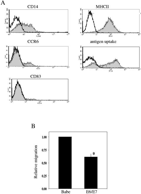
FIG. 2.
Characterization and migration of LPLCs. (A) LPLCs were generated from CD14+ peripheral blood monocytes as described in Materials and Methods. On day 6, LPLCs were stained with anti-CD14, CCR6, CD83, and major histocompatibility class II monoclonal antibodies or incubated with conjugated ovalbumin and were analyzed using a FACScan instrument and CellQuest software. Open histograms indicate cell staining with the isotype-matched negative-control antibody. (B) Primary human foreskin keratinocytes stably expressing vector alone (Babe) or HPV-16 E6/E7 (E6/E7) were treated with Poly(I) · Poly(C). Following treatment, cells were washed with PBS and refed with minimal media for 4 h. Culture supernatants were collected and used in multiwell chamber assays. Values are expressed as migration of LPLCs in response to E6/E7 supernatant relative to migration in response to supernatant from control cells (Babe). Results are expressed as the means ± SD of three experiments using three independent cell lines (* P < 0.001).

