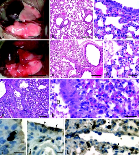
FIG. 2.
Pathologies of mice intranasally inoculated with 106 PFU of rescued parental Tx/91, 1918 HA/NA:Tx/91, or 1918 HA/NA:WSN recombinant influenza viruses. Gross photographs (a and d) and photomicrographs (b, c, and e through h) of hematoxylin and eosin-stained tissue sections are shown. (a) Lungs of a mouse inoculated with Tx/91 virus; lungs at day 5 p.i. appear normal except for a small focus of pneumonia in left lung lobe. Bar = 5 mm. (b) Mild lymphocytic peribronchitis with mild alveolitis in a mouse inoculated with Tx/91; day 5 p.i. Bar = 200 μm. (c) Predominant peribronchial histiocytic alveolitis with iatrogenic atelectasis in a mouse inoculated with Tx/91; day 5 p.i. Bar = 25 μm. (d) Large areas of pneumonia affecting most of the left lung and cardiac lobe of the right lung. Smaller areas of pneumonia in apical and azygous lobes of the right lung of a mouse inoculated with 1918 HA/NA:Tx/91; day 5 p.i. Bar = 5 mm. (e) Diffuse severe alveolitis in a mouse inoculated with 1918 HA/NA:Tx/91; day 5 p.i. Bar = 200 μm. (f) Severe neutrophilic-to-histiocytic alveolitis adjacent to bronchiole with necrotizing bronchitis. Furthermore, some alveoli have pulmonary edema evident as protein crescents in a mouse inoculated with 1918 HA/NA:Tx/91; day 5 p.i. Bar = 20 μm. (g) Severe necrotizing bronchitis with severe alveolitis in a mouse inoculated with 1918 HA/NA:WSN; day 5 p.i. Bar = 100 μm. (h) Higher magnification of panel g showing prominent necrotic debris in bronchial lumen (labeled “B” within panel) and abundant neutrophils and histiocytes in alveoli. Bar = 20 μm. (j) Two virus-positive bronchial epithelia (arrowheads) in a mouse inoculated with 1918 HA/NA:Tx/91; day 5 p.i. Bar = 10 μm. (k) Virus-positive AMs (arrowheads) in a mouse inoculated with 1918 HA/NA:Tx/91; day 5 p.i. Bar = 10 μm.

