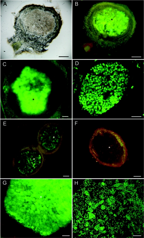
FIG. 6.
Light microscopy of fresh nodules of Mimosa spp. inoculated with gfp-tagged or wild-type (WT) Burkholderia strains Br3461 (A to D) and MAP3-5 (E to H). A Mimosa pudica nodule infected with Br3461-gfp was viewed with transmitted light (A) or epifluorescence (B). Panels C and D show a Mimosa bimucronata nodule infected with Br3461-gfp viewed under epifluorescence (C) and an infected cell from the same nodule containing fluorescent bacteroids viewed using CLSM (D). (E and F) Mimosa pudica nodules infected with MAP3-5-gfp (E) or MAP3-5 WT (F) viewed under epifluorescence. Note that the nodule containing WT MAP3-5 does not fluoresce (F). (G) Mimosa pigra nodule infected with MAP3-5-gfp viewed with epifluorescence. Note that the infected zone fluoresces intensely green (*), but also that the meristematic region has red fluorescence, possibly due to the presence of polyphenolic compounds. (H) CLSM of fluorescent bacteroids within an infected cell from an M. pigra nodule infected with MAP3-5-gfp. Bars, 100 μm (A, B, C, E, and F), 5 μm (D), 50 μm (G), and 10 μm (H).

