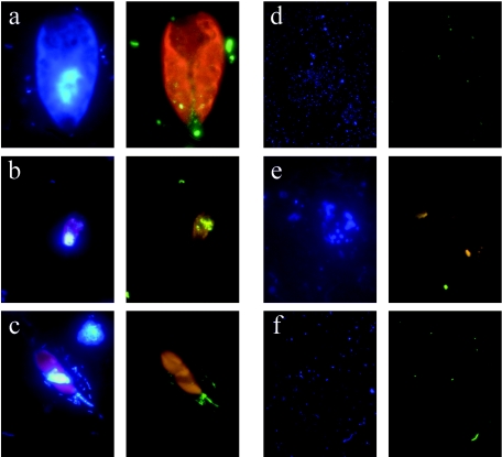
FIG. 1.
Epifluorescence microscope photographs. Within each lettered panel, the left image depicts DAPI staining, and the right one depicts CARD-FISH staining with EUB338 probe on Cryptomonas ovata (right image using filter set 24) (a), EUB338 probe on Rhodomonas minuta (b), ARCH915 probe on Dinobryon cylindricum (c), EUB338 probe after G fixation (d), ARCH915 after LFT fixation and K permeabilization showing cell fragments of unidentified phytoplankton (e), and ARCH915 probe after LFT fixation and LA permeabilization (f).

