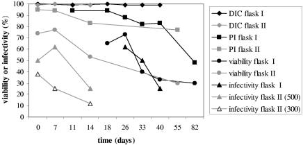
FIG. 1.
Ageing of C. parvum oocysts in sterile surface water at 15°C. Differential interference contrast microscopy (DIC), propidium iodide exclusion without acid treatment (PI), and the percentage of oocysts that both excluded PI after acid treatment and contained sporozoites (viability) indicate oocyst viability. Oocyst infectivity is indicated by the percentage of infected HCT-8 cell monolayers (infectivity). Flask I contained approximately 1,100 oocysts · 100 μl−1, and flask II contained approximately 500 oocysts · 100 μl−1. The results for a dilution containing approximately 300 oocysts · 100 μl−1 are also shown.

