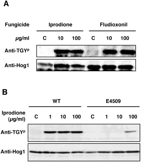
FIG. 3.
Phosphorylation of the C. heterostrophus BmHog1p MAPK by iprodione and fludioxonil. (A) BmHog1p phosphorylation by iprodione and fludioxonil in the wild-type strain. Mycelia of HITO7711 (WT) were incubated in CM with 10 or 100 μg/ml iprodione or fludioxonil for 10 min. Prepared mycelia were incubated in CM with ethanol and dimethyl sulfoxide as a control (indicated by C). The cells were harvested, and total protein extracts were prepared as described in Materials and Methods. Protein samples (50 μg) were subjected to SDS-PAGE and blotted onto nitrocellulose membranes for Western blot analysis. Phosphorylated BmHog1p was detected using anti-dually phosphorylated p38 antibody (indicated by anti-TGYp). The total amount of BmHog1p was measured using anti-Hog1 C terminus antibody (indicated by anti-Hog1). (B) BmHog1p phosphorylation by iprodione in the wild-type strain and the dic1-deficient strain. Mycelia of HITO7711 (WT) and E4509 were used for analysis. Prepared mycelia were incubated in CM with 1, 10, or 100 μg/ml iprodione for 10 min. Prepared mycelia of the strain tested were incubated in CM without iprodione as a control (indicated by C). The procedures for Western blot analysis were the same as those described above.

