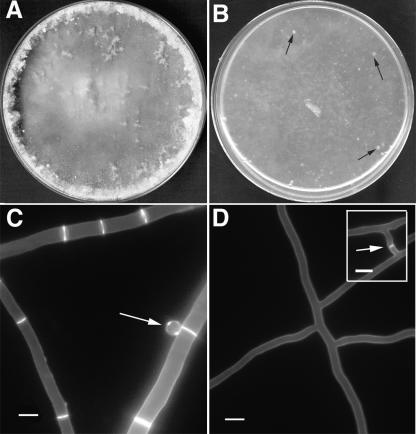
FIG. 2.
Phenotype of an N. crassa rho-4 mutant. (A) Wild-type N. crassa hyphae and conidia after 5 days of growth on Vogel's MM plates. (B) A rho-4 mutant on Vogel's MM plates forms only thin mycelia with patches of leaking cytoplasm (black arrows) and no conidia. (C) Wild-type N. crassa hyphae stained with calcofluor (1 μg/ml) show bright staining at septa and cell walls. An arrow points to a conidium. (D) rho-4 mutant hyphae stained with calcofluor (1 μg/ml) show cell wall staining, but no septal staining. (Inset) rho-4 mutants undergo cell fusion. Arrow marks area of recent cell fusion event. Calcofluor staining does not show a septum, but the intervening cell wall of the fusion hyphae, which is degraded upon completion of hyphal fusion. Bar, 10 μm.

