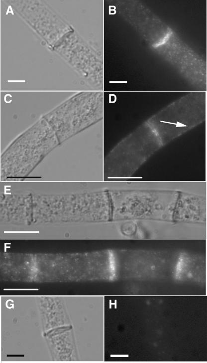
FIG. 4.
Immunofluorescence microscopy of HA-tagged RHO-4. (A) Differential interference contrast (DIC) image of CR21-12 (his-3+::Pgpd-HA-rho-4; rho-4) treated with anti-HA antibodies. (B) Fluorescent micrograph of the same hypha as in panel A. (C) A second DIC micrograph of CR21-12 treated with anti-HA antibodies. (D) The same hypha as that shown in panel C, showing both plasma membrane localization (indicated by an arrow) and septal localization of HA-RHO-4. (E) DIC micrograph of CR21-12 treated with anti-HA antibodies showing multiple septa. (F) Fluorescent micrograph of same hypha as in panel E showing that all septa are labeled. (G) DIC micrograph of FGSC 988 (wild-type strain) treated with anti-HA antibodies. (H) Fluorescent micrograph of the same hypha in panel E shows little nonspecific antibody binding. Bars, 10 μm.

