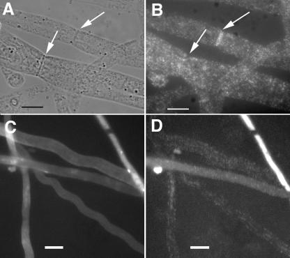
FIG. 5.
Actin localization in wild-type and rho-4 strains. (A) DIC image of wild-type (FGSC 988) hyphae treated with anti-actin antibodies. Arrows show septa. (B) Fluorescent micrograph of hyphae in panel A. Arrows are the same as in panel A. Note absence of actin staining in the lower septa. (C) Calcofluor-stained image of hyphae from rho-4 mutant CR7-7 treated with anti-actin antibodies. Hyphae were stained with calcofluor to show absence of septa in rho-4 mutants. (D) Fluorescent micrograph of hyphae shown in panel C. Bar, 10 μm.

