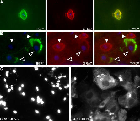
Figure 5. The Morphological Changes of the IIGP1 Accumulations at the PV Are Accompanied by Loss of T. gondii GRA7 from the PV and its Dissemination throughout the Cytoplasm.
(A and B) IFN-γ-induced astrocytes were infected with T. gondii for 2 h (A) or 6 h (B) and stained for IIGP1 (green) and GRA7 (red) (filled arrowheads: IIGP1-negative PVs, open arrowheads: IIGP1-positive PVs). Nuclei were stained with DAPI. (C) Uninduced (left) and IFN-γ-induced (right) astrocytes were infected with T. gondii and stained for GRA7 at 4 h post-infection. Exposure conditions for the two images were the same.

