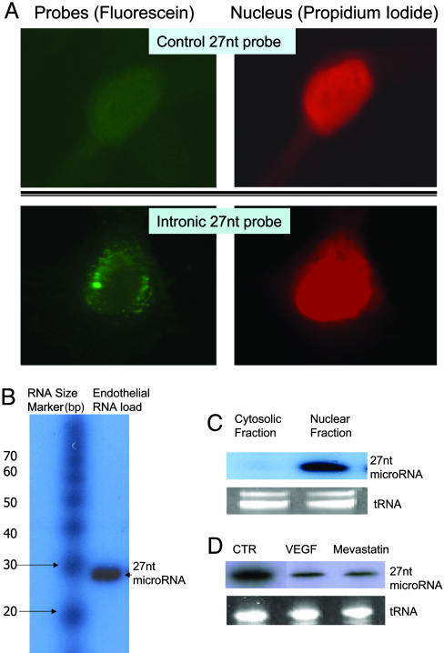
Fig. 6.
Presence of endogenous 27-nt microRNA in cultured HAECs. (A) In situ hybridization was performed in cultured HAECs. HAECs were grown up to 50% confluence. After fixation and dehydration, cells were incubated in hybridization buffer containing the DIG-labeled 27-nt oligo-probe at 42°C for 16 h. The anti-DIG-fluorescein (dilution of 1:500 in blocking solution) was then applied to the cells at room temperature for 45 min. After counterstaining the nuclei with propidium iodide, samples were visualized under fluorescence microscopy. The fluorescein (green) showed that the 27-nt RNA was distributed within the nucleus, and in the perinuclear area of the HAECs. The staining appears to be in a punctate form. No obvious fluorescein signal was detectable in HAECs when control probe (27-nt single strand DNA complementary to luciferase gene) was used. (B) Northern blotting for the 27-nt microRNA from the endothelial small RNA extracts. The sizes of small RNA markers (Ambion) are labeled on the left. The band at the position approximate to 27-nt was hybridized to the [γ-32P]-labeled 27-nt oligo-probe is indicated on the right. (C) Northern blotting shows that the 27-nt microRNA is mainly located in the nuclear fraction. The cytosolic fraction contains none or only trace of the 27-nt microRNA. (D) A representative Northern blot of three separate experiments for the total cellular 27-nt microRNA from the endothelial cells treated by 10 ng/ml VEGF or 10 μM mevastatin for 24 h during culture. Both VEGF and mevastatin reduced 27-nt microRNA significantly (48.2 ± 11.1% and 39.6 ± 7.9% of the levels in the control cells, respectively).

