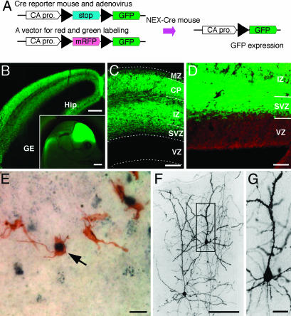
Fig. 1.
Distribution of GFP-positive cells marked by NEX-Cre mediated cell lineage analysis. (A) Schematic diagram of cell lineage tracing using double-transgenic (NEX-GFP) mice. In NEX-positive cells, Cre recombinase removes a loxP-flanked stop sequence, bringing together the chicken actin (CA) promoter and the coding region for GFP. (B) Frontal section of the NEX-GFP mouse at E15. Subcortical regions, i.e., ganglionic eminence (GE), lack GFP staining (Inset, sagittal view of the E15 embryonic head). (C) Frontal section of the NEX-GFP neocortex at E15. Note that GFP-positive cells are absent from the VZ and MZ. (D) A clear boundary emerges between VZ and SVZ after counter-staining with propidium iodide. (E-G) One day after injection of recombinant adenovirus into the lateral ventricle of NEX-Cre embryos (E14), a number of cells in the SVZ are immunostained for both GFP (brown) and P-H3 (dark blue) (arrow in E). Three weeks after injection, basically all NEX-GFP-marked cells developed into pyramidal neurons in the upper cortical layers (E). At higher magnification, NEX-GFP-marked cells (boxed in E) reveal spiny processes (F). (Scale bars: 1 mm in B Inset; 200 μm in B; 100 μm in C and F; 50 μm in D; and 20 μm in E and G.) Sections in B-D are 20 μm, and those in E-G are 50 μm in thickness.

