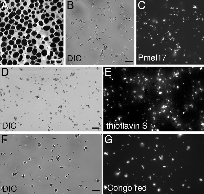
Figure 1. Purified Melanosomes Stain with Amyloidophilic Dyes.
Melanosomes were isolated from bovine RPE and choroid and visualized using transmission electron microscopy (A; scale bar = 1 μm), differential interference contrast microscopy (DIC) (B, D, and F; scale bars = 10 μm), indirect immunofluorescence using a Pmel17-specific antibody (C), or the thioflavin S (E) or Congo red (G) amyloidophilic fluorophores. Images (B) and (C), (D) and (E), and (F) and (G) are paired.

