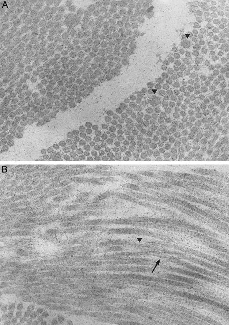
Figure 1 .
Ultrastructural findings in the skin of patient 2. A, On the transverse section (28,400×), the collagen fibrils show a distinct variability in diameter; some fibrils are larger and irregular in outline (arrowhead). B, On the longitudinal section (28,400×), the deposition of granulofilamentous material along the collagen fibrils is visible (arrowhead). In some foci, collagen fibrils have an unraveled and disorganized aspect (arrow).

