
Figure 1.
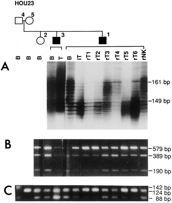
Testing of DNA samples from family HOU23, including members HOU23-04 (unaffected father), HOU23-05 (unaffected mother), HOU23-02 (unaffected child), HOU23-03 (affected child), and HOU23-01 (affected child), as depicted on pedigree shown above gels. The family represents an example of germline mosaicism, details of which are reported elsewhere (Rose et al. 1999). Individual lanes are labeled to indicate tissue from which DNA was extracted: B = blood lymphocytes, T = AML sample from HOU23-03, lT = left kidney AML, rT1–rT6 = six individual right-kidney AML samples, and rNK = right-kidney normal tissue from HOU23-01. Decrease in intensity of bands indicating loss of the wild-type TSC2 allele in some cells of the AMLs from patient HOU23-01 are observed in A–C. A, Marker D16S525 is located ∼95 kb telomeric to the 5′ end of TSC2. Both HOU23-01 and HOU23-03 are heterozygous, with alleles of 149 bp and 161 bp present. The 161-bp allele was significantly reduced in the lT, rT1, rT2, rT5, and rT6 of HOU23-01, whereas the 149-bp and 161-bp alleles were of equal intensity in rT3 or rT4 of HOU23-01 and in the AML (T) from HOU23-03. B, Patients were informative for the EcoRV polymorphism in exon 40 of TSC2, with heterozygotes showing three bands at 579, 389, and 190 bp, respectively. Decrease in the intensity of the wild-type allele was found in the same AML DNAs (lT, rT1, rT2, rT5, and rT6) as in A, indicating LOH for the wild-type allele in some cells from which the DNA was extracted. C, The marker D16S665 is located ∼75 kb from the 3′ end of TSC2. The father (HOU23-04) is heterozygous for the marker, with alleles of 142 and 88 bp, and the mother is homozygous for the 124-bp allele. The results showed the same five AMLs (lT, rT1, rT2, rT5, and rT6) with decreased intensity of the paternal 88-bp allele, while retaining the maternal 124-bp allele. The paternal 142-bp allele is not inherited by any of the offspring in the family.