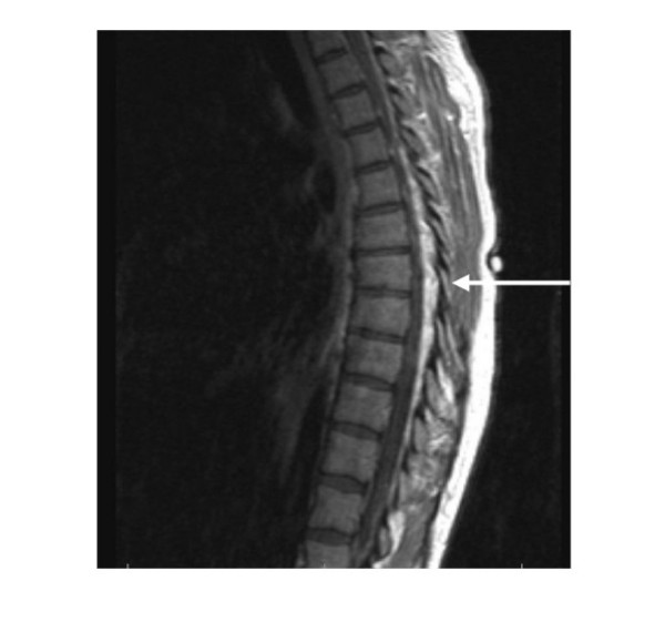
Figure 1.
multi-planar, multi-sequence MRI imaging of the thoracic spine. Pre and post contrast sagittal and axial T1, and turbo T2 sequences acquired. An irregular masslike density predominantly T1 and T2 hyperintense with a serpiginous contour is demonstrated in epidural location, as a mass surrounding the thoracic cord. The lesion extends from the T1 level distally through the lower thoracic spine.

