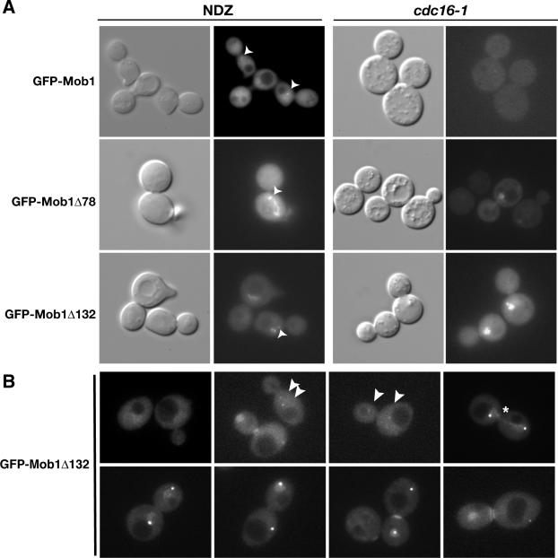
Figure 2.
Dynamics of truncated Mob1p localization. (A) Mob1Δ78 and Mob1Δ132 localize to nuclei in metaphase-arrested cells. GFP-Mob1p, GFP-Mob1Δ78, and GFP-Mob1Δ132 were analyzed in nocodazole (NDZ)-treated wild-type cells (left panels) and conditional cdc16-1 cells (right panels). The cells were synchronized in metaphase, as described in the text. The strains used in the NDZ experiments were FLY316, FLY1066, and FLY1147. The cdc16-1 cells (FLY547) contained pRS314-GFP-Mob1, pRS314-GFP-Mob1Δ78, or pRS314-GFP-Mob1Δ132. We obtained similar results for cdc16-1 cells expressing integrated Mob1p (unpublished data). (B) Representative images of GFP-Mob1Δ132 in homozygous diploid cells (FLY1875) from various stages of the cell cycle. The arrowheads point to GFP-Mob1Δ132 on early mitotic SPBs. Note that the second SPB in the last image on the bottom panel is out of focal plane. The pattern of GFP-Mob1Δ78 fluorescence was similar to GFP-Mob1Δ132 (Supplementary Movies 2–4). See Results for detailed description. All images in B are merges of 6 × 0.2-μm Z-sections.

