Figure 5.
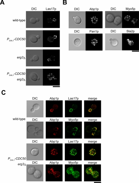
The cdc50Δ erg3Δ mutant displays cytoplasmic mislocalization of actin patch components. (A) Localization of Las17p-EGFP patches in the cdc50Δ erg3Δ mutant. The Las17p-EGFP-expressing strains examined were KKT108 (wild-type), KKT109 (PGAL1 -3HA-CDC50), KKT110 (erg3Δ), and KKT111 (PGAL1-3HA-CDC50 erg3Δ). Cells were cultured in SDAW at 30°C for 12 h, followed by confocal microscopic observation. Central focal plane images are shown. (B) Localization of other actin patch components in the cdc50Δ erg3Δ mutant. The strains examined were PGAL1-3HA-CDC50 erg3Δ mutant expressing Abp1p-EGFP (KKT158), Myo5p-EGFP (KKT90), Pan1p-EGFP (KKT163), or Sla2p-EGFP (KKT219). Cells were cultured and images were acquired as in A. (C) Colocalization of Abp1p-mRFP1 with Las17p-EGFP and Myo5p-EGFP in the cdc50Δ erg3Δ mutant. The strains examined were KKT234 (wild-type) and KKT237 (PGAL1 -3HA-CDC50 erg3Δ) expressing both Las17p-EGFP and Abp1p-mRFP1, and KKT229 (wild-type) and KKT232 (PGAL1 Δ) -3HA-CDC50 erg3 expressing both Myo5p-EGFP and Abp1p-mRFP1. Cells were cultured and images were acquired as in A. Bars, 5 μm.

