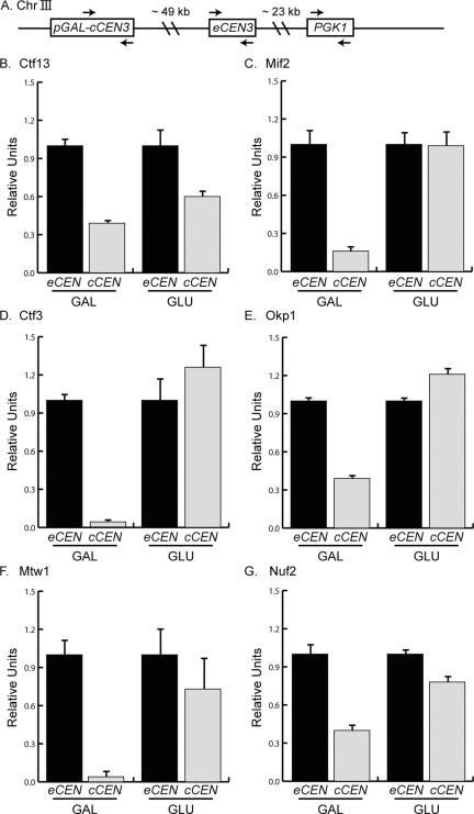
Figure 1.
Kinetochore proteins from each subcomplex can assemble in G1. (A). Schematic of chromosome III (Chr III) indicates the position of the cCEN (pGAL-cCEN3) and the negative control locus (PGK1) with respect to the eCEN (eCEN3). Arrows indicate the PCR primer sets used to detect these loci in ChIP experiments shown in the subsequent figures. (B–G). Strains expressing Ctf13-myc13, Mif2-myc13, Ctf3-myc13, Okp1-myc13, Mtw1-myc13, and Nuf2-myc13 were arrested in G1 with αF and then transferred to media containing GAL or GLU. ChIP was performed with anti-myc antibody. Relative units indicate the eCEN:PGK1 ratio or the cCEN:PGK1 ratio and are normalized to an eCEN:PGK1 value of 1.0 for each experiment. All kinetochore proteins tested localize to the active cCEN in G1.

