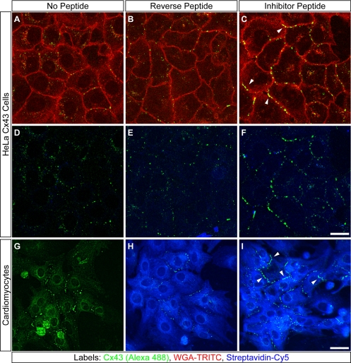
Figure 5.
Peptide inhibition of Cx43–ZO-1 interaction increases the extent of membrane-localized Cx43 GJ plaques in HeLa cells and cardiomyocytes. Confocal optical sections of HeLa Cx43 cells (A–F) and neonatal cardiomyocytes (G–I) cultured for 72 or 24 h, respectively, without peptide (A, D, and G), or with reverse (B, E, and H) or inhibitor peptide (C, F, and I). All cells were immunolabeled for Cx43 (A–I). Peptides were detected with streptavidin-Cy5 (D–I). WGA-TRITC delineates HeLa cell membranes (A–C). Arrowheads (C and I) denote accumulation of membrane-localized Cx43 plaques in inhibitor-treated cells. Bars, 20 μm (A–F); 80 μm (G–I).

