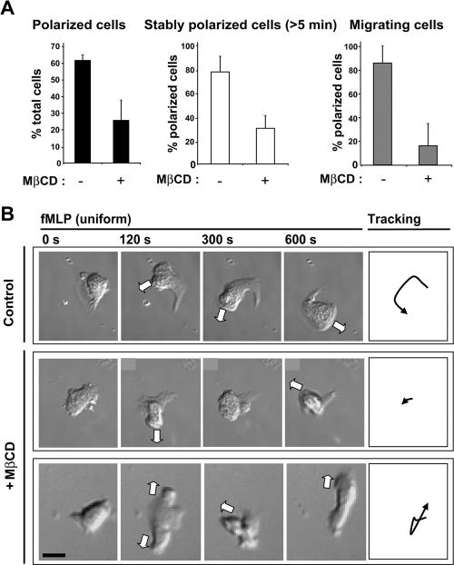
Figure 1.
Controlled cholesterol depletion disturbs polarization and migration induced by uniform fMLP-stimulation. (A) Graphs showing: (left) the percentage of control and MβCD-treated cells able to polarize upon stimulation with fMLP (100 nM, 14 min); middle, the percentage of polarized control or MβCD-treated cells able to maintain their polarity for at least 5 min; right, the percentage of polarized control and MβCD-treated cells that undergo migration over a 10-min time course. Results are means ± SD for four separate experiments (n = 126 control cells and n = 146 cholesterol-depleted cells). (B) Time-lapse DIC images of individual cells stimulated with fMLP. Top row, a stably polarized control cell; middle and bottom rows, MβCD-treated cells forming multiple protrusions, either successively (middle) or simultaneously (bottom). The white arrows indicate the directions of the protrusions. Right, trajectories of the center of each cell. Scale bar, 10 μm.

