Figure 1.
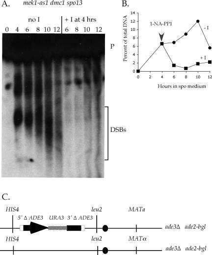
Double-strand breaks in a mek1-as1 dmc1 spo13 diploid in the absence of inhibitor and after addition of inhibitor after 4 h in spo medium. (A) The mek1-as1 dmc1 spo13 diploid, NH574::pNH251, was transferred to sporulation medium and incubated at 30°C for 4 h. At that time, 1 μM 1-NA-PP1 was added to one-half of the sporulating cells and returned to the incubator. At 2-h intervals, cells were fixed and analyzed for DSB formation at the YCR048w hotspot as described in Woltering et al. (2000). Numbers above each lane indicate hours after transfer to sporulation medium. The bracket indicates the DSB fragments. P, parental band. (B) Quantitation of the DSBs shown in A. (C) Configuration of the sister chromatid recombination reporter present on chromosome III in the isogenic spo13 (NH567::pNH250) and mek1-as1 dmc1 spo13 (NH574::pNH251) diploids (Kadyk and Hartwell, 1992). The black oval indicates the centromere on chromosome III. The white box indicates the region of shared homology between the 5′Δ ADE3 and 3′Δ ADE3 truncations between which recombination can occur.

