
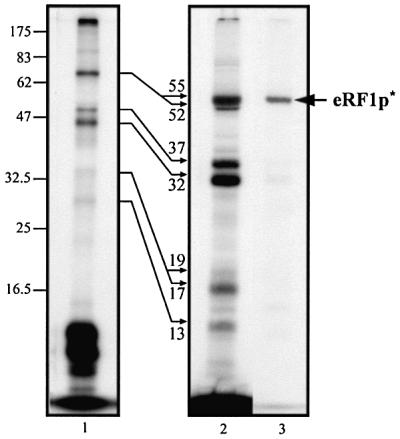
Fig. 5. Crosslink patterns obtained with internally labeled s4U*GA mRNA analog and phased ribosomes in the presence of eRF1: lane 1, without nuclease treatment; lane 2, after microccocal digestion of tRNAs. Reaction conditions: 0.1 µM mRNA, 0.2 µM ribosome and, where indicated, 2 µM tRNAAsp and 6 µM eRF1. Autoradiogram after 12.5% SDS–PAGE. To localize the His6-tagged eRF1p* product (arrow), an aliquot of the mixture obtained after irradiation of the complete mixture was nuclease treated and passed through an Ni-NTA column. The fraction eluted with 150 mM imidazole was analyzed in parallel (lane 3). Molecular masses of the rPp* and eRF1p* obtained after nuclease digestion are indicated (in kDa) close to the thin arrows.