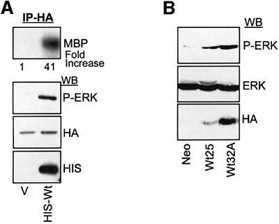
Fig. 4. IEX-1 potentiates ERK activity. (A) CHO cells were co-transfected with 1 µg of pcDNA-HA-ERK1 and 1 µg of either empty vector (V) or plasmid encoding His-Wt-IEX, as indicated. Twenty-four hours later, ERK activity was measured in anti-HA immunoprecipitates (IP) by western blotting or kinase assay using MBP as a substrate. (B) Stable clones of UT7 cells expressing different levels of HA-IEX-1 or control cells (Neo) were stimulated for 30 min with TPO and ERK activation was measured by western blotting with anti-phosphoERK antibodies.

An official website of the United States government
Here's how you know
Official websites use .gov
A
.gov website belongs to an official
government organization in the United States.
Secure .gov websites use HTTPS
A lock (
) or https:// means you've safely
connected to the .gov website. Share sensitive
information only on official, secure websites.
