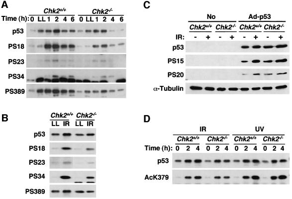
Fig. 7. Phosphorylation and acetylation of p53 protein in Chk2–/– cells after IR. (A) ES cells were exposed to 10 Gy IR or treated with 10 µM of LLnL (LL) and then lysed at the indicated times after treatment. Immunoprecipitates prepared with PAb421 were subjected to immunoblot analysis with the p53-specific antibodies PAb421 and PAb240 (p53) or with antibodies specific for p53 phosphorylated on the indicated serine residues (PS18, PS23, PS34 or PS389). (B) Similar amounts of p53 protein from irradiated or LLnL (LL)-treated ES cells harvested 2 or 4 h after treatment, respectively, were subjected to immunoblot analysis of p53 as in (A). (C) Chk2+/+ and Chk2–/– MEFs infected with or without adenovirus-expressing human p53 were cultured for 24 h, the cells were exposed (or not) to 10 Gy IR and then lysed 2 h after IR. Total cell lysates were analyzed by immunoblot using either the human p53-specific monoclonal antibodiy DO-1 (p53), phosphoserine-specific antibodies [anti-human p53 PabSer(P)15 (PS15) and anti-PAb(P)20 (PS20)] or anti-α-tubulin as probes. (D) Differentiated ES cells were exposed to IR (10 Gy) or UV light (50 J/m2) and lysed at the indicated times after treatment. Immunoprecipitates prepared with PAb421 antibodies were subject to immunoblot analysis with p53-specific PAb421 and PAb240 or with antibodies specific for p53 acetylated on lysine379 (AcK379).

An official website of the United States government
Here's how you know
Official websites use .gov
A
.gov website belongs to an official
government organization in the United States.
Secure .gov websites use HTTPS
A lock (
) or https:// means you've safely
connected to the .gov website. Share sensitive
information only on official, secure websites.
